Where we are today
In recent years, research efforts have increasingly focused on unravelling the complex biological processes underlying CLN3 Batten disease, including the role of CLN3 dysfunction in lysosomal activity, autophagy, and neuroinflammation. This research has revealed important clues and direction for the development of several promising therapeutic avenues.
INVESTIGATIONAL THERAPEUTIC APPROACHES
Gene Therapies
Advances in gene-based therapies for CLN3 disease aim to address the underlying genetic cause by restoring or compensating for the function of the defective CLN3 gene. Approaches include gene replacement therapy, where functional copies of the gene are delivered using adeno-associated virus (AAV) vectors, and antisense oligonucleotides (ASOs) which seek to modify RNA splicing and improve the production of functional CLN3 protein. In addition, gene editing techniques like CRISPR/Cas9, which seek to correct mutations at the DNA level are currently being explored in preclinical (cell and animal) models.
These therapies hold promise for slowing disease progression by preventing neuronal loss and reducing neuroinflammation. However, challenges remain, including safe and efficient gene delivery to the brain, immune responses to treatment, and the need for early intervention before significant neurodegeneration occurs. Ongoing preclinical and clinical trials are working to refine these approaches and determine their long-term effectiveness in treating CLN3 disease.
Drug Repurposing
Drug repurposing means finding new ways to use medicines that are already approved for other conditions. Because these drugs have already been tested for safety, this approach can help speed up the search for effective treatments for Batten disease. Previously, mycophenolate an immunosuppressive drug, and flupirtine, a non-opioid analgesic have been explored as potential treatments for CLN3 disease. However, in the absence of convincing data, this research has not advanced.
More recently, miglustat, a glucosylceramide synthase inhibitor, has been explored as a potential treatment for CLN3 disease due to its ability to reduce glycosphingolipid accumulation, a contributor of CLN3 disease pathology. Approved in various markets for treatment of other lysosomal storage disorders Gaucher and Niemann-Pick type C diseases, miglustat is now the subject of ongoing clinical studies currently sponsored by the Beyond Batten Disease Foundation in partnership with THX Pharma (formerly Theranexus).
Further information on the miglustat (Batten-1) program, including published studies, can be found here. [https://beyondbatten.org/research/bbdf101/]
CURRENT TREATMENT OPTIONS
At present, there is no approved treatment or cure for CLN3 disease. Standard of care remains limited to supportive and palliative care and includes:
- Symptom Management: Use of antiepileptic drugs to control seizures, medications to alleviate movement disorders such as dystonia, and interventions to manage behavioral and psychiatric symptoms.
- Multidisciplinary Support: Holistic multidisciplinary approach to care includes physical, occupational, nutritional and speech therapies to help maximize and maintain mobility, independence and quality of life.
- Palliative Care: Focused on optimizing overall well-being and providing supportive care for the affected individual and their family, and may be engaged as early as time of diagnosis (not only in the mid-later stages of disease).
In summary, although current treatment options remain largely supportive, significant progress in understanding CLN3 pathology is paving the way for innovative therapies that may potentially slow or halt the disease progression in the future.
February 2026 Research Update from Dr. Ineka Whiteman, BBDF Principal Scientist
BBDF is proud to have co-authored three recent publications. Summaries and links to these papers are below.
A timeline of symptom onset and disease progression in CLN3 disease
Whiteman, I.T., Cook, A.L., Augustine, E.F. et al. Orphanet J Rare Dis (2026).
We’re excited to share the latest review on the natural history of CLN3 disease – a valuable resource for affected families, healthcare providers and researchers.
CLN3 disease is one of the most prevalent forms of Batten disease, usually beginning in the first decade of life and progressing in severity, with life expectancy in the 20’s or 30’s. Despite two decades of natural history studies, there has been no clearly defined timeline describing when key symptoms appear and how the disease progresses, making it difficult to plan optimal care and design effective clinical trials.
Inspired by conversations with families and clinicians, and led by BBDF’s Dr Ineka Whiteman together with investigators from the original natural history studies, this comprehensive meta-analysis brings together data from over 400 affected individuals to map the onset and progression of 13 core symptoms of classical CLN3 disease. The findings reveal a clear chronological pattern, beginning with early vision loss and advancing through cognitive, behavioural and motor decline, cardiac manifestations, feeding difficulties and end of life.
By defining a clearer disease trajectory, this study offers practical anticipatory guidance for clinicians and families. It also highlights the need for standardised longitudinal natural history data to improve care and support future therapeutic development for CLN3 disease.
Read the article here.
Limited therapeutic efficacy of N-acetyl-L-leucine in a mouse model of CLN1 disease
Ziółkowska, E.A., Pagán Torres, N.A., Chen, H. et al. Sci Rep 16, 3033 (2026).
N-acetyl-L-leucine (NALL) is an oral compound with reported neuroprotective, anti-inflammatory and metabolic effects, recently approved for treatment of the lysosomal storage disorder Niemann-Pick disease type C. Researchers at Washington University in St. Louis tested this agent in a mouse model of CLN1 disease to see whether NALL as a monotherapy (single therapy) could alleviate symptoms. Treatment was started either before symptoms appeared or after symptoms had begun and continued through advanced disease stages. The results showed that NALL alone did not meaningfully slow disease progression, improve overall motor function, reduce brain inflammation, or extend survival in this model, although a modest, quantifiable benefit was observed on locomotor (walking) stability when treatment began early.
These findings, although overall non-significant, provide important information in understanding the underlying biology of CLN1 disease. Moreover, as the authors state, “these negative results should not be interpreted as a dismissal of NALL’s therapeutic potential in general but rather as a demonstration of its limited value as a monotherapy in CLN1 disease.”
Future research may be well directed toward testing NALL in combination therapy, in other NCL subtypes, or both.
This study was led by Dr Ewa Ziółkowska and Dr Jonathan Cooper, co-designed and co-authored by our Principal Scientist, Dr Ineka Whiteman and supported by the BBDF.
Enteric nervous system degeneration in human and murine CLN3 disease, is ameliorated by gene therapy in mice
Ziółkowska, E.A., Williams, L.L., Jansen, M.J. et al. Acta neuropathol commun 13, 260 (2025).
This ground-breaking study in human and murine CLN3 disease provides first evidence of direct effects of CLN3 disease on GI dysfunction, treated with gene therapy in CLN3 mice.
Individuals with CLN3 disease frequently experience severe gastrointestinal (GI) problems that can significantly reduce quality of life and may contribute to death. This study shows a substantial loss of neurons and glia in the enteric nervous system – the master regulator of the bowel function – leading to bowel distension and impaired gut function. Importantly, AAV-mediated CLN3 gene therapy delivered systemically in CLN3-deficient mice prevented much of this nerve damage and preserved bowel function later in disease. This preclinical study provides early evidence that gene therapy may help address GI complications by targeting the underlying cause, expanding the field beyond our traditional understanding of Batten disease as a disorder of the central nervous system (CNS) only.
This study also highlights the profound impact of patient organ donation. Examining bowel samples from individuals affected by Batten disease enables researchers to confirm that the disease effects observed in animal models is present, and therefore clinically relevant, in human patients. The generosity of families who choose to donate tissue makes it possible to study the disease directly, validate animal models, and move the field forward in meaningful ways.
Read more.
Summer 2025 Research Update from Dr. Ineka Whiteman, BBDF Principal Scientist
Impact of CLN3 Disease on Child Quality of Life and Family Function.
Vermilion J, Augustine EF, Mink JW, McDermott MP, Vierhile A, Pereira-Freitas M, Adams HR.
Pediatr Neurol. 2025 Jun 16;170:17-25. doi: 10.1016/j.pediatrneurol.2025.06.008. Online ahead of print.
Summary: A new study from the University of Rochester Batten Center shows that children with CLN3 disease have significantly reduced quality of life, which worsens as the disease progresses. While families report a substantial overall impact, this does not appear to change in line with symptom severity, highlighting the ongoing need for targeted support for both patients and caregivers.
The Wechsler intelligence scale for children, fourth and fifth editions perform comparably in children with Batten disease.
Adams HR, Augustine EF, Bonifacio K, Collins A, Vierhile AE, Mink JW. Orphanet J Rare Dis. 2025 Aug 7;20(1):413. doi: 10.1186/s13023-025-03923-w.
Summary: Researchers at the University of Rochester Batten Center found that two versions of a common cognitive test, the WISC-IV and WISC-V, produce comparable results in children and young adults with Batten disease. This means data from both versions can be combined to better understand the cognitive decline associated with the disease over time.
Other publication highlights:
| Natural history and variants in neuronal ceroid lipofuscinoses: Uncoupling genotype and phenotype.
Mole SE. Dev Med Child Neurol. 2025 Aug 2. doi: 10.1111/dmcn.16442. Online ahead of print.
Ziółkowska EA, Jablonka-Shariff A, Williams LL, Jansen MJ, Wang SH, Eultgen EM, Wood MD, Hunter DA, Sharma J, Sardiello M, Reese R, Pestronk A, Sands MS, Snyder- Warwick AK, Cooper JD.
|
April 2025 Research Update from Dr. Ineka Whiteman, BBDF Principal Scientist
Keep up to date with the latest clinical trial and natural history study news with our Clinical Studies Chart on the BDSRA Foundation website. Check it out here.
CLN3 Alcyone Therapeutics
CLN-301 Gene therapy (AAV9; CNS)
NCT03770572 Ph1/2 – active, not recruiting (USA)
Formerly licenced to Amicus Therapeutics (until February 2024).
*APRIL 2025 UPDATE* Alcyone Therapeutics Advances Pipeline for CLN-301 Gene Therapy for CLN3 Batten Disease
Alcyone Therapeutics Advances Pipeline for CLN-301 Gene Therapy for Treatment of CLN3 Disease
Alcyone Therapeutics has announced strategic financing to advance therapeutic candidate ‘CLN-301’ in an expanded collaboration with Nationwide Children’s Hospital, Columbus, OH.
CLN-301 is a potential first-in-class AAV9 gene therapy in Phase 1/2 clinical development for CLN3 Batten disease. Formerly licensed to Amicus Therapeutics before being returned to Nationwide in January 2024, the investigational gene therapy has been in clinical trial since 2018, with four individuals treated and followed for over 5 years (see https://clinicaltrials.gov/study/NCT03770572).
As stated in Alcyone’s recent press release, “initial results from a Phase 1/2 trial with CLN-301 demonstrated safety and therapeutic effects in a cohort of four patients compared to the natural progression of CLN3 Batten disease. On the Unified Batten Disease Rating Scale, patients in the CLN-301 study remained stable with a slope of -0.22 points per year over three years. By comparison, an increase of 2.86 points per year, leading to an overall 8.6-point increase in impairment over 36 months would be typical according to natural history data. Typical outcomes for patients with CLN3 Batten disease include rapid vision loss, cognitive and motor decline, and behavioral issues. The majority of patients treated with CLN-301 and followed for over five years to date have shown maintenance of skills in motor and cognitive function and overall health compared to natural history data.”
Read Alcyone’s full announcement here.
February/March 2025 Research Update from Dr. Ineka Whiteman, BBDF Principal Scientist
Batten Disease Global Research Initiative Inaugural Grants Announced!
Starting with our biggest news this month, we were thrilled to announce the winners of the inaugural Batten Disease Global Research Initiative (BDGRI) Research Grants. In our press release on March 18, we were proud to reveal the recipients, awarding a total of USD $320,000 to six groundbreaking projects worldwide.
Read the full announcement here.
BBDF is proud to be a Funding Partner and support John Ostergaard’s and Sara Mole’s projects. More information can be found here.
Rare Disease Week – February 24-26
I had the privilege of participating in Rare Disease Week on Capitol Hill, Washington DC. Hosted by the EveryLife Foundation and Rare Disease Legislative Advocates, this annual event unites advocates from across the country to make their voices heard by their Members of Congress. Throughout the week, participants heard from leaders in the field on policy proposals impacting the rare disease community, including:
- Support for steady and robust leadership, federal biomedical research funding, and public health agencies.
- Reauthorization of the Rare Pediatric Disease Priority Review Voucher Program.
- Inclusion of the Accelerating Kids’ Access to Care Act (AKACA).
Right now, Congress is juggling three separate but intertwined legislative processes related to funding government operations, each of which have direct implications for these policies: the Continuing Resolution, the Fiscal Year (FY) 2026 Appropriations Cycle, and Budget Reconciliation. The EveryLife Foundation has provided a brief overview to help breakdown what is happening, what is at stake, and what can be done to support the resources and services that drive our rare disease research, therapy development, and access to care. You can read the review here:
How Congressional Budget Decisions Shape Rare Disease Funding—And What You Can Do
Click here to check out the highlights from our Week on Capitol Hill.
Highlights from the 2025 Gordon Research Conference on Lysosomal Diseases – March 1-7, Pomona, CA
Held every two years, Gordon Research Conference (GRC) is focused on cutting-edge and unpublished research. There is always a wonderful turnout from the Batten disease research community (pictured below) and I thoroughly enjoy the opportunity to connect with so many throughout the week-long meeting.
The GRC is designed to foster in-depth discussion, new collaborations and to strengthen existing collaborations among attendees from clinical, academic and biotech sectors. Key topics this year included novel gene replacement and gene-editing approaches, biomarker discovery, and cellular mechanisms driving disease progression. Sessions delved into how lysosomal function is impaired in various diseases and how this knowledge can inform broader applications in biology and medicine. Emerging areas such as the role of lysosomes in neurodegeneration, energy metabolism, infection, and immunity were also explored.
I was honored to have presented a poster on some of our recent collaborative research titled CLN3 Batten disease: A Timeline of Symptom Onset and Disease Progression, and was also invited to present on a Career Mentorship panel, as part of the Gordon Research Seminar (GRS), a satellite meeting to GRC for young researchers. I enjoyed the opportunity to share my experiences in academic research, the biotech industry and now in the patient advocacy space, highlighting how my role in Batten disease patient advocacy has by far been the most rewarding of these. I hope I may have inspired some of our young researchers to consider this career pathway sometime in their futures!
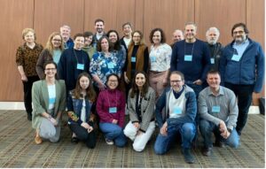
Image: Batten disease researchers, clinicians and patient advocates from around the world came together at the GRC in California in March.
EVENTS
MEET-THE-EXPERT WEBINAR – Research update & Communication Strategies in CLN2 & CLN3 Batten disease
On April 2nd, BDSRA Australia hosted a live webinar event with Speech Pathologists Lottie Morison and Prof. Angela Morgan from The Centre of Research Excellence – Translational Centre for Speech and Disorders at Murdoch Children’s Research Institute (MCRI) Melbourne. They shared the research paper ‘Speech, Language and Non-verbal Communication in CLN2 and CLN3 Batten Disease’, which was recently published in the Journal of Inherited Metabolic Disease. As a co-author on the paper, BBDF is proud to support this important research.
In the webinar, presenters provided an overview of the collaborative research, including practical strategies to support communication for individuals with CLN2 and CLN3 disease, such as tailored speech and language intervention, and augmentative and alternative communication (AAC).
A recording of this webinar, and additional speech, language and communication resources are now available. Please email Nikki Hopkins at info@bdsraaustralia.org for further information.
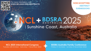
NCL2025 Congress ABSTRACTS and REGISTRATION NOW OPEN!
The 19th International Congress on Neuronal Ceroid Lipofuscinosis (NCL) will be held form October 28th – November 1st, 2025, in the stunning surrounds of the Sunshine Coast, Queensland, Australia. As the premier conference on Batten disease, the bi-annual NCL Congress brings together world-leading researchers, healthcare providers, industry partners, innovators and patient advocacy leaders from around the globe.
Abstracts and Early-Bird Registrations are now open! Abstracts must be submitted by June 6th.
We hope to see you Down Under! Visit www.NCL2025.org for more details.
As always, thank you for your support and dedication to our shared mission.
Warm regards,

January 2025 Research Update from Dr. Ineka Whiteman, BBDF Principal Scientist
The Power of Family Involvement in Batten Disease Research
In Batten disease research, as with other rare diseases, true progress is best achieved when patients and families are at the center of the conversation. Their lived experiences provide critical insights that drive meaningful advancements in science, clinical care, and drug development.
Family perspectives help inspire research ideas and shape study designs, ensuring they reflect real-world challenges and priorities; patients and their families offer invaluable data through participation in natural history studies, biobanking, and through involvement in clinical trials.
But beyond the science, patient and family involvement keeps research grounded in its true purpose: improving lives. By fostering strong collaborations between researchers, clinicians, and the Batten disease community, together we create solutions that are not just innovative but also practical and impactful.
This month’s Research Highlights, both of which I was honored to have been involved, are outstanding examples of how family involvement can make a profound difference in the fight against Batten disease. On behalf of the research teams represented in these two highlighted articles, I want to extend our deepest gratitude to all the families who have contributed to this important work. Whether through participation in speech and language assessments, sharing insights with researchers, or generous tissue donation, your dedication and generosity helps bring hope to countless others. Thank you for being an essential part of this journey.
Research Highlights
New research on speech and language in CLN2 and CLN3 disease
Progressive speech and language impairment is a prominent feature of Batten Disease, yet to date there has been no systematic characterization of speech and language in this condition.
Understanding the decline in communication in Batten disease helps us better understand the disease biology, may be an important measure of treatment efficacy in clinical trials, and critically, can help inform families and clinicians optimize speech & language therapy approaches.
In this study, led by Prof. Angela Morgan and PhD candidate Lottie Morison at the Murdoch Children’s Research Institute (MCRI) in Melbourne Australia, the team sought to understand the speech and language features, support needs and strengths in the two most common forms of Batten disease: CLN2 and CLN3 disease.
The results of this research, published in the Journal of Inherited Metabolic Disease demonstrate (1) the importance of clinical education and awareness that speech and language difficulties can be early signs of Batten disease; (2) tailored speech and language therapies are important to support speech and language skills, especially training communication partners; and (3) many people with Batten disease would benefit from early Augmentative and Alternative Communication (AAC) access to support communication for as long as possible.
This paper is freely available to read here Speech, Language and Non‐verbal Communication in CLN2 and CLN3 Batten Disease
Free Patient Resources are also available, including Plain Language Summary of this research and Fact Sheets about speech & language in CLN2 and CLN3 Batten disease. Thanks to the Translational Centre for Speech Disorders at MCRI.
Download the PDF resources here: Genes – Centre of Research Excellence in Speech and Language
NCL2025 Congress website now live!
Beyond Batten Disease Foundation is thrilled to be a main sponsor for the 19th International Congress on Neuronal Ceroid Lipofuscinosis (NCL) in Queensland, Australia. As the premier conference on Batten disease, the bi-annual NCL Congress brings together world-leading researchers, healthcare providers, industry partners, innovators and patient advocacy leaders from around the globe.
In 2025, we are excited to support an invaluable opportunity for meaningful engagement and shared learning between Congress delegates and the Batten disease family community.
The website is now live – visit NCL 2025. Here you can register to stay up to date with latest meeting information and news including venue and accommodation details, programs and abstract submissions.
We hope to see you Down Under!
As always, thank you for your support and dedication to our shared mission.
Warm regards,
Characterisation of sleep in a mouse model of CLN3 disease revealed sex-specific sleep disturbances.
Kane KM, Iradukunda D, McLouth CJ, Guo LZ, Wang J, Subramoniam A, Huffman D, Donohue KD, O’Hara BF, Sunderam S, Wang QJ.
J Sleep Res. 2025 Jan 28:e14461. doi: 10.1111/jsr.14461. Online ahead of print.
PMID: 39873354
Zhao JQ, Feng BY, Ye ZL, Ma XY, Du JZ, Li JM, Wu WL, Gao JJ, Li SJ, Peng SY, Huai JS, Ge LH, Lu CB.
Acta Pharmacol Sin. 2025 Feb;46(2):338-352. doi: 10.1038/s41401-024-01377-7. Epub 2024 Sep 16. PMID: 39284877
Speech, Language and Non-verbal Communication in CLN2 and CLN3 Batten Disease.
Morison LD, Whiteman IT, Vogel AP, Tilbrook L, Fahey MC, Braden R, Bredebusch J, Hildebrand MS, Scheffer IE, Morgan AT.
J Inherit Metab Dis. 2025 Jan;48(1):e12838. doi: 10.1002/jimd.12838. PMID: 39821609
Open-label evaluation of oral trehalose in patients with neuronal ceroid lipofuscinoses.
Della Vecchia S, Gammaldi N, Ricca I, Mero S, Doccini S, Ardissone A, Bagnoli S, Battini R, Colombi E, Favaro J, Furlan R, Giordano L, Ingannato A, Mandelli A, Manzoni FMP, Milito G, Moroni I, Nacmias B, Nardocci N, Parmeggiani L, Pezzini F, Pietrafusa N, Sartori S, Specchio N, Trivisano M, Ets ACL, Simonati A, Santorelli FM; A-NCL ETS Group.
J Neurol. 2025 Jan 7;272(1):94. doi: 10.1007/s00415-024-12790-7. PMID: 39775944
The use of nanocarriers in treating Batten disease: A systematic review.
Henke L, Ghorbani A, Mole SE.
Int J Pharm. 2024 Dec 16:125094. doi: 10.1016/j.ijpharm.2024.125094. Online ahead of print. PMID: 39694161 Review.
Dwojak E, O’Mard D, Zou J, Wassif CA, Burkett S, Eckhaus M, Rueda Faucz F, Padilla C, Villasmil R, Zheng W, Dang Do AN.
Stem Cell Res. 2024 Dec;81:103563. doi: 10.1016/j.scr.2024.103563. Epub 2024 Sep 18. PMID: 39317061
Zhang D, Xu F, Bao Y, Xu Y.
Zhonghua Yi Xue Yi Chuan Xue Za Zhi. 2024 Dec 10;41(12):1469-1472. doi: 10.3760/cma.j.cn511374-20240927-00510.
PMID: 39653353 Chinese.
Vacuolated lymphocytes: a diagnostic biomarker for CLN3-related Batten disease.
Cruz-Pimentel M, Parameswarappa DC, Ryu G, Klatt R, Vincent A.
Can J Ophthalmol. 2025 Mar 20:S0008-4182(25)00114-0. doi: 10.1016/j.jcjo.2025.02.021. Online ahead of print.
Enzyme Replacement Therapy in CLN2-Associated Retinopathy.
Priglinger C, Courage C, Maier EM.
Klin Monbl Augenheilkd. 2025 Mar;242(3):213-218. doi: 10.1055/a-2528-7886. Epub 2025 Mar 24. PMID: 40127655 Review.
Tagless LysoIP for immunoaffinity enrichment of native lysosomes from clinical samples.
Saarela D, Lis P, Gomes S, Nirujogi RS, Dong W, Rawat E, Glendinning S, Zeneviciute K, Bagnoli E, Fasimoye R, Lin C, Nyame K, Boros FA, Zunke F, Lamoliatte F, Elshani S, Jaconelli M, Jans JJ, Huisman MA, Posern C, Westermann LM, Schulz A, van Hasselt PM, Alessi DR, Abu-Remaileh M, Sammler EM.
J Clin Invest. 2024 Dec 26;135(4):e183592. doi: 10.1172/JCI183592. PMID: 39724071; PMCID: PMC11827837.
Priglinger CS, Courage C, Lotz-Havla AS, Gerhardt M, Ehrt O, Kurz M, Pudritz H, Rudolph G, Jackson CB, Maier EM.
Neuropediatrics. 2025 Apr;56(2):142-146. doi: 10.1055/a-2510-5592. Epub 2025 Jan 7. PMID: 39776429
TRAM-LAG1-CLN8 family proteins are acyltransferases regulating phospholipid composition.
Sheokand PK, James AM, Jenkins B, K Lysyganicz P, Lacabanne D, King MS, Kunji ERS, Siniossoglou S, Koulman A, Murphy MP, Petkevicius K.**
Sci Adv. 2025 Feb 21;11(8):eadr3723. doi: 10.1126/sciadv.adr3723. Epub 2025 Feb 19.
PMID: 39970228
** 2025 Batten Disease Global Research Initiative Grant Recipient
Shilatifard A, Ben-Sahra I.
J Clin Invest. 2025 Feb 17;135(4):e188507. doi: 10.1172/JCI188507.
PMID: 39959975
Yu Z, Yan J, Liu Z, Wang H, Luo G, Chen H.
Front Cell Dev Biol. 2025 Jan 23;13:1508714. doi: 10.3389/fcell.2025.1508714. eCollection 2025.
PMID: 39917569 Free PMC article.
Neuronal Ceroid Lipofuscinosis-Concepts, Classification, and Avenues for Therapy.
Zhang Y, Du B, Zou M, Peng B, Rao Y.
CNS Neurosci Ther. 2025 Feb;31(2):e70261. doi: 10.1111/cns.70261.
PMID: 39925015 Review.